DERMOID SINUS IN THE RHODESIAN RIDGEBACK
(A Review by a Veterinarian)
Taken from South African Rhodesian Ridgeback Club, Ridgeback Review
Surgery of a Dermoid Sinus
Detection of a Dermoid Sinus on the tailset
THE FORMATION AND SIGNIFICANCE OF DERMOID SINUS
The Rhodesian Ridgeback is a modern breed of dog that originated in the late nineteenth century, by the
crossing of indigenous Hottentot dogs with various European breeds introduced into the Cape by the
early settlers.
The breed standard was established with the formation of the Rhodesian Ridgeback Club of Bulawayo in
1922. The main characteristic of the breed is, as its name implies - a ridged back, which is formed in the
haircoat along the top midline of the dog's back. The ridge is formed by hair, which grows in the
opposite direction to the hair of the surrounding coat.
Breeders of Ridgebacks are aware of a well-known defect which occurs in the breed, the Ridgeback
"Cyst" or as it is more correctly named in the scientific terminology, the Dermoid Sinus. (Dermoid -
arising from the skin, Sinus - a cavity or channel).
Dermoid Sinuses are narrow tube-like structures, which are derived from a skin defect. They penetrate
from the skin surface to varying depths downward into the muscles and towards the spinal cord. They
are situated in the midline of the neck and croup, which is in front and behind the area occupied by the
ridge (Fig 1).
Fig 1: Areas marked X indicate the sites at which dermoid sinuses may develop.
This is the only known congenital defect that occurs regularly in the breed. (Congenital means that the
defect is formed before birth). When considered as a defect in the dog family as a whole, Dermoid
Sinuses occur only very rarely in dogs, other than Ridgebacks or Crossbred Ridgebacks. It must
therefore be obvious that it is an inherited defect which has become widespread in the "blood lines" of
the breed as a result of the early selective breeding of the original stock from which the Ridgebacks of
today have been produced.
The incidence of the defect throughout the breed is not known, as the recording of the numbers of
Dermoid Sinus affected pups in litters has not been done on a scale large enough to enable a statistical
analysis to be carried out. In fact, the occurrence of Dermoid Sinus affected pups in the litters of
breeders has been kept confidential, as most breeders feel that there is considerable stigma attached to
dogs and bitches amongst whose offspring Dermoid Sinus affected puppies occur.
At this point I would like to state that with the present situation of breeding with selected outstanding
dogs and bitches, no breeder without a program of progeny testing can be sure that his "blood line" is
free from the hereditary Dermoid Sinus. (The hereditary aspects of the condition will be dealt with in
part two of this article). Thus, every purchased Ridgeback may be considered a potential carrier of the
condition.
THE FORMATION OF DERMOID SINUS
To understand the way, in which a Dermoid Sinus is formed, it is necessary to have some idea of how the
embryo develops from a single fertilised egg cell in the womb of the bitch. Dermoid Sinus is a congenital
defect that arises from a defect in the development of the embryo of a puppy.
A fertilised egg resulting from a successful mating is a single simple cell. From this cell a puppy
consisting of millions of specialised cells, which constitute the tissues and organs, must be formed in 63
days.
Fig 2: Early stages of cell division.
A : Single cell of fertilised egg.
B : 2 cell stage.
C : 4 cell stage.
D : 8 cell stage.
E : Multi-celled Spherical Mass, many cell divisions later.
This process in accomplished by a rapid increase in the number of cells by cell division. The fertilised
egg (a single cell) divides into two cells and subsequent divisions each double the previous number of
cells, so that the numbers very rapidly increase. In the ten successive divisions, 1042 cells are produced,
and it can be seen that, by this means, the total number of cells is soon very large.
The next stage is the organisation of the mass of cells produced to form a puppy. The organisation
process, which takes place for about the first three weeks of pregnancy, is called the embryonic
development. When the embryo is fully developed, a complete miniature puppy is formed which now
becomes known as a foetus. The next six weeks of pregnancy only results in the increase in size of the
foetus to its normal birth size.
Dermoid Sinuses arise from a defect in the development of the embryo. Cell division gives rise to a
spherical mass of cells. The outer layer of these cells will eventually become the skin of the puppy.
Another part of the body also develops from this outer layer of cells. This is the brain and the spinal
cord, which runs from the head to the base of the tail. The problem now is - how does the outer layer of
cells give rise to the brain and the spinal cord?
This is accomplished by the formation of a long groove over half the surface of the spherical mass of
cells. The groove deepens and then its outer edges close together giving rise to a tube-like structure.
This tube-like structure which is later to become the brain and spinal cord, sinks deeper below the
surface layer and becomes detached from it. This process is shown in Figure 3.
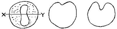
Fig 3: Schematic Formation of Brain and Spinal Cord
Formation of groove on
surface of sphere of cells as
seen from above groove.
Section through X-Y to show
outer layer of cells folding
inwards.
Deepening of the fold.
Closing over of edges
of the groove.
Closed.
Separation of outer layer
(skin) from tubular structure
(spinal cord).
Dermoid Sinuses occur when small areas of attachment between the outer layer of cells (the skin) and
the tubular structure (later to become the brain and the spinal cord) remain.
In the puppy this defective separation of the embryological tissues is present as a thin tubular
attachment extending from the skin of the midline of the top surface of the dog to the deeper tissues
below, and as deep as the spinal cord in some cases.
The depth to which this tubular skin defect penetrates is the criterion used for the classification of
four types of Demioid Sinuses, shown in Figure 4 below.
Fig 4: Cross section through a dog's neck. Types of Dermoid Sinuses
Types of Dermoid Sinuses
TYPE I
Penetrates below the skin surface, its fatty tissue overlying the neck muscles.
TYPE II
Penetrates into the muscles of the neck.
TYPE III
Penetrates to the supraspinous ligament, which runs over the top of the vertebrae.
TYPE IV
Penetrates to the spinal cord between the vertebrae.
THE SIGNIFICANCE OF DERMOID SINUS
The detrimental effects of Dermoid Sinus are not just concerned with the fact that a visible anatomical
defect is apparent in affected animals, but rather the complications which can arise as a result of a
Dermoid Sinus becoming infected with bacteria.
The narrow tube of skin which descends below the skin surface is lined with all the normal skin
structures and of special significance are : hair; sweat; and oil glands.
The thin central cavity, which runs down the Dermoid Sinus, becomes filled in time with hair, skin oil and
skin scales. The contents usually become an ideal medium where bacteria, which are normally present on
the skin, may grow. They gain access to the material through the small pore-like opening at the point of
attachment of the Dermoid Sinus on the skin surface.
The accumulated skin secretion undergoes a process of putrefaction and the skin barrier of the Sinus
walls breaks down and bacteria invade the tissues deep below the skin surface. This usually results in
the formation of an abscess, which eventually ruptures to the outside and drains as a chronically
discharging purulent wound.
Extensive surgical and medical treatment may be necessary to clear up such a complication and in some
cases septic dermoid sinus may be unresponsive to treatment.
If a Dermoid Sinus is recognised in a dog before it becomes septic, it can be removed surgically, with a
good chance that no further complications will occur. In most cases, however, owners of animals are not
aware of the presence of a Dermoid Sinus and shortly thereafter sepsis almost always sets in.
Subsequently, the owners are obliged to obtain veterinary treatment to resolve the distressing
complications. This may be costly to the dog owner and embarrassing to the breeder when it is pointed
out that he has sold a dog with a latent defect.
DIAGNOSIS OF DERMOID SINUS
In the puppy, Dermoid Sinus can be detected by raising the skin in a longitudinal fold along the top
midline in the area in which Dermoid Sinuses are known to occur (i.e. in front of and behind the ridge).
If the skin fold is raised with one hand and the skin allowed to slip back and forward between the thumb
and forefinger of the other hand the presence of the Sinus can be felt as a thin cord-like structure
between the two layers of skin (Fig 5)
Figure 5: Diagnosis of Dermoid Sinus
Feeling for presence of Dermoid Sinus by sliding longitudinal fold of skin between index finger and
thumb.
Raising the skin fold in this way tenses the tissues and a Dermoid Sinus will be pulled tautly between its
skin attachment in the top midline and its attachment in the muscles below.
The diagnosis can be confirmed by shaving the hair from the skin over the point at which the Dermoid
Sinus is attached. A small pore like opening in the skin from which a small tuft of hair protrudes is
usually seen. This is the opening of the Dermoid Sinus on the skin surface. The older the puppy, the
thicker the Sinus will be and the more easily it may be recognised.
It must be realised, however, that the recognition of a Dermoid Sinus in puppies may not always be as
easy a procedure as the above description may suggest. If it is missed, a Dermoid Sinus may lie dormant
for years before it comes to the notice of an owner by becoming septic. If in doubt the professional
assistance of a Veterinarian should be obtained.
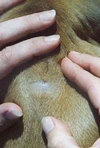
Next pictures shows how to find a Dermoid Sinus in a puppy. This pics have been taken by Anke Terbruggen.
You can see a suspicious spot, where the hairs seem te have a slightly different colour and structure.
When you feel the spot, there seems te be a small "lump" under the skin.
When the skin is lifted, the DS is clearly visible, as a "thread" which runs down from the skin to the spinal cord.
Also note the "dip" in the skin.
Next step is to shave the area, then the hole is clearly visible.
Look, this puppy has two holes.
On this other one, there is only a hole.
Now that you know the puppy has a Dermoid Sinus you have to decide what to do, to cull the pup or remove the Dermoid
Sinus with surgery. If you decide the last one, you will have to spay/neuter the puppy.